
Calcifications in the hip tendons – Types, causes, treatment

There are three main types of calcification that can form in the tendons around the hip, and each requires slightly different treatment. Read on to understand what type you might have and what the best treatment choices are.
Remember, if you need help with an injury, you're welcome to consult one of our physios online via video call.

The terms tendinitis, tendonitis, tendinosis, and tendinopathy mean the same thing for all practical purposes, and we use these interchangeably in our articles. Read more (opens in new window).
In this article:
Which hip tendons can develop calcifications?
You can develop calcifications in any of the tendons that surround the hip joint – it can sometimes even form in the muscles. The most commonly affected are the gluteal tendons (glute med, glute max, and glute min) in the area of the greater trochanter (the nobbly bone on the outside of the hip).

However, calcifications can also develop in the proximal (upper) hamstring tendons, adductor tendons (inner groin), and proximal rectus femoris (upper front thigh muscle) at the front of the hip.

Types of tendon calcification
Hard calcifications associated with chronic tendinopathy
These usually show up as small specs or lumps of hard calcification in a tendon that has chronic or ongoing tendinopathy or tendonitis. The calcifications are thought to form when calcium phosphate salts leak out of the injured tendon cells.
But not everyone with chronic tendonitis develops these calcifications, and there is evidence that other aspects of your general health (diabetes, hypothyroidism, etc.) might actually play a role in triggering this process.
In one study where researchers reviewed the scans of 202 people with chronic gluteal tendinopathy (defined as having symptoms for more than three months), they found that 36% of them also had calcifications. There’s no similar data for how common this is in the proximal hamstring or adductor tendons.

Soft calcifications associated with calcific tendonitis
These calcifications show up as large lumps on scans and are made up of large quantities of carbonated hydroxyapatite - researchers describe it as having a soft chalky texture, similar to that of toothpaste.
These pockets of soft calcification are formed when some of your tendon cells change into cartilage-type cells, which then start producing large quantities of carbonated hydroxyapatite.
Once again, we’re not yet 100% sure what causes this to happen – some researchers suspect it might be triggered by an injury, but there is evidence that certain factors in your general health (diabetes or being overweight), hormone changes, and inflammatory arthritis might trigger it.
When you have this type of calcification, your doctor will likely diagnose you with calcific tendonitis of the hip, although that term is often also (incorrectly) used for the other types discussed in this article.

Interestingly, with calcific tendonitis, researchers often report that the rest of the tendon looks healthy and uninjured on scans (no signs of chronic tendonitis or tendinopathy).
However, I did find a couple of case reports by surgeons who found large quantities of soft calcium in the proximal hamstring tendons during surgery to repair tendon tears and also a case where there were signs of gluteal tendinopathy. So, it might sometimes accompany other tendon injuries.
Calcific tendonitis usually follows a five-stage process:
Precalcific stage: Some of the cells change from tendon cells to cartilage-type cells.
Formative stage: The calcium deposits form – this is usually a pain-free process but can sometimes cause a dull ache.
Resting stage: The calcium then just sits there for a while (resting stage) and, because it usually doesn’t cause pain, you might not even know it’s there.
Resorption stage: This is usually the most painful time and starts when the body notices the calcium and sends clean-up cells (inflammation) and creates new blood vessels in the tendon to help remove it.
Finally, the tendon repairs with the correct type of cells.
👉 Sometimes the process gets stuck in Stage 3 or 4, and then there are specific treatments that might help to move it along – more on this later.
Bony spurs (enthesophytes)
These show up as little bony bumps or points in the area where your hip muscle tendons attach into the bone. The reason we included them in this article is because they start off as calcium deposits that then turn into bone.
The area where a tendon or ligament attaches into a bone is called the enthesis. That area contains cartilage cells, which, under normal conditions, helps with ongoing bone repair. Bone spurs develop when the cartilage cells in one area of the enthesis become very active, creating excessive amounts of calcium, which then turn into bone.

Researchers suspect that the body creates these spurs in an attempt to reinforce that area – the argument being that when the tendon pulls excessively on that part of the bone, it causes it to become overworked and the cartilage cells creates the extra bone in an attempt to prevent injury.
Bone spurs are often found in tendons that also show signs of tendinopathy or tendonitis, a condition that is also caused by overuse. But not always – many people with bone spurs don’t get any pain from them and don’t even know they’re there.


Best scans to diagnose hip calcifications
Using a combination of X-rays and ultrasound scans usually provides the most complete picture.
The X-rays are pretty good at showing all three types of calcification, while the ultrasound scan provides a bit more detail about the exact size of the calcification and tendon health, e.g. whether there is a tendon tear or tendonitis.
💡 If you had to do only one scan, the ultrasound would come out as the top choice.
MRI scans are actually not that great at showing calcifications. Large calcifications can look very similar to tendon tears on MRI scans, and surgeons have been surprised to find large quantities of soft calcification during surgery that did not show up on these scans.

How much of your pain is down to the calcification vs. other factors?
The tricky thing to get one's head around is that, even though these calcifications or bone spurs might “look” on scans as if they should be painful, they often don’t cause any trouble.
Hard calcifications and bony spurs usually don’t cause pain unless they are very big, break up (fractures), or you also have tendinopathy or tendonitis at the same time – and then the pain usually settles as the tendonitis heals despite the calcification or spur remaining, which suggests that the pain might actually not have been caused by the calcification.
In chronic Achilles tendonitis that doesn’t respond to regular treatment, surgeons will often shave the injured part of the tendon away and also remove the calcifications, which then helps the pain settle – but then they find that the calcifications return after a few months, despite the patient still not having any pain. This also points to the tendon injury likely being the source of the pain rather than the hard calcification.
Soft calcifications act differently. Yes, they can often be pain-free while forming, but they might cause an annoying dull ache if they become very large. Once you enter the resorption stage, the pain is usually quite intense, and this is when most patients first become aware of it and go to see a doctor.
💡 The good news is that this very painful period usually only lasts around three weeks in shoulders – I couldn’t find evidence for how long it takes in hips.
How to treat the different types of hip calcification
Hard calcifications and bone spurs
Because the evidence suggests that the pain and symptoms are not coming from the calcification, but rather the accompanying tendon injury, the first line of treatment is focused on getting the tendonitis or tear to recover. If, however, this fails to produce results, surgery is an option.
The treatment steps will look slightly different depending on your specific case, but they usually follow this type of pattern:
Relative rest – This means adapting or reducing your daily activities and exercise to a level that your tendon is currently happy and able to tolerate. This is crucial to allow the pain to settle and recovery to take place.
A well-structured rehab plan – The aim is to stimulate the tendon through exercises to rebuild and repair the injured area. It’s important that the exercises you do match your tendon’s current strength and sensitivity, otherwise they can make things worse.
Other treatments – If you are stuck and you’re not making progress with the above, then other treatments like shockwave or injections might also be useful.
Surgery – In some cases, pain might persist despite you doing everything right, and then surgery can be effective. The type of surgery will depend on what the surgeon feels is most appropriate in your case.
👉 You can find detailed advice on how to best treat tendonitis or tendinopathy in specific tendons and adapt it to your specific case here:
💡 Shockwave therapy might help to improve the symptoms of the tendonitis (but not too early in treatment), but it can’t break up hard calcifications or help your body to absorb it.

Treatment for soft calcifications (calcific tendonitis)
Here, the treatment is slightly different because:
the body will often absorb the calcifications by itself
when the calcification enters the resorption stage, it usually causes a huge inflammatory response (which is not the case in chronic tendonitis).
If you´re in the very painful resorption stage, treatments are usually aimed at reducing excessive load on the tendon and getting the inflammation under control:
Anti-inflammatory medication might help.
Applying ice for short periods to the injured area can be useful (10min on, take off for 10min, reapply ice for 10min).
Relative rest – You don´t have to rest completely; just limit activities to what you can do without significantly increasing your symptoms.
Strong exercises should be avoided during this period.
👍 This approach has been shown to work well for calcific tendonitis around the hip (even when very severe).
If your calcification is “stuck” in a specific stage and isn’t being absorbed, the following treatments might help it move along:
Focused shockwave has been shown to help break up this type of calcification and get the body to absorb it.
Barbotage and Lavage are techniques (which are often combined) where the doctor will use a needle to puncture the soft calcium deposit, repeatedly stabbing it while injecting clean water to help dissolve it before drawing it out.
Corticosteroid injections are often used in shoulders to help the very acute pain to settle. There is a small risk that these injections might also affect tendon health and it is currently thought that it might be best to only use it in cases that do not want to respond to other treatments.
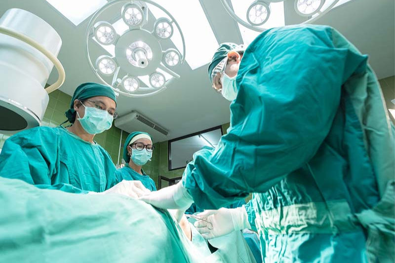
If your pain persists for more than six months, despite trying all of the other treatments mentioned above or the calcification is very large, surgery might be an option and does seem to produce good results.
💡 With soft calcifications, rehab exercises are usually only started once the acute pain has started to settle a bit.
However, in this case, the exercises are not strictly needed to address the tendon injury (unless you also have a tendinopathy or tear) but rather to restore the strength and endurance you lost during the period of inactivity due to the pain you experienced.
At this point, the exact same rehab principles then apply here as for general tendon rehab:
💡 Regardless of the type of calcification you have, it’s important that the rehab exercises must match your tendon’s current strength and sensitivity – if they’re too intense, they’ll just irritate your tendon and increase your symptoms.
How we can help
Need help with an injury? You’re welcome to consult one of the physio team at SIP online via video call for an assessment of your injury and a tailored treatment plan.

We're all UK Chartered Physiotherapists with Master’s Degrees related to Sports & Exercise Medicine or at least 10 years' experience in the field. But at Sports Injury Physio we don't just value qualifications; all of us also have a wealth of experience working with athletes across a broad variety of sports, ranging from recreationally active people to professional athletes. You can meet the team here.


About the author
Maryke Louw is a chartered physiotherapist with more than 20 years' experience and a Master’s Degree in Sports Injury Management. Follow her on LinkedIn and ResearchGate.


